- About Us
- Ilizarov Technique
- Process
- Case Studies
Fractures
• Acute Fracture - Comminuted Fracture Distal Humerus - Compound Fracture & VAC • Nonunion - Proximal Tibia - Tibia - Distal Humerus • Malunion - Femur Bone Deformity
• Acute Correction
- LRS - Bilateral Tibia - LRS - Rotational Deformity Femur(with Shortening) - LRS - Oblique Plane Deformity Femur - Nailing • Ilizarov Correction - Proximal Tibia - Distal Femur • Hexapod Correction - Metaphyseal Dysplasia Bone Infection
• Infection - Femur • Infected Nonunion - Proximal Femur Joint Problems (Arthritis)
• Hip Replacement • Knee Replacement • Intra Articular Osteotomy • High Tibial Osteotomy (HTO) • Arthrolysis - Elbow • Ilizarov Hip Reconstruction - FAQs
- Testimonials
News & Updates


Nailing - Valgus Deformity Distal Femur
 |
||
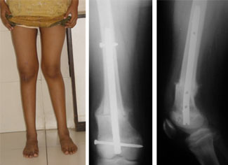
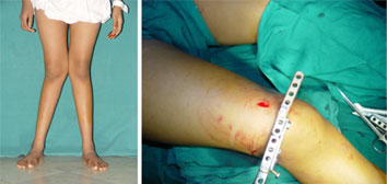
| The picture on the left side shows an example of bilateral valgus deformity(knock knees) at the distal femur i.e. reduced angle at the outer side of the thigh bone close to the knee. Here the growth if the patient is complete and there is no limb length discrepancy. The picture on the right side was taken during the surgery. The metallic contraption is used as a guide for cutting the bone. The incision shown is about 1.25 cm long. The bone is cut using this incision and with minimal soft tissue stripping, which helps in better bone healing and better results for the patient. |
||
| Percutaneous Osteotomy | ||
 |
||
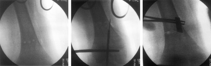
| Percutaneous osteotomy is performed using minimally invasive technique through a small incision, just enough to permit the osteotome with minimal soft tissue stripping. The pictures here show the xrays done during surgery. Drill holes are made using the jig. The entry path for the nail in the distal part of the bone is marked using k-wire and cannulated drill bit. Another instrument like osteotome is used to mark the correct angle. The holes are then connected and osteotomy is completed using the osteotome. |
||
| Translation, Fixation | ||
 |
||
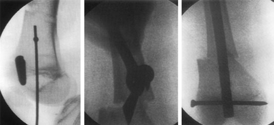
| These are x-ray pictures taken during surgery. The left one shows the path of entry of the guide wire as seen from the side. The picture in the center shows how the bone is translated(shifted on the long axis) at the osteotomy site. This is done in accordance with osteotomy rule 2(i.e when the osteotomy is away from the CORA-center of rotation axis.) The picture on the right shows the final picture after the insertion of the intra-medullary nail in the corrected position with locking bolt holding the nail to the bone. |
||
| Healing | ||
|
||
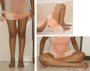
| Final Result | ||
|
||