| |
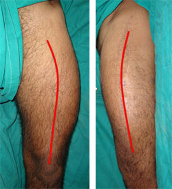 |
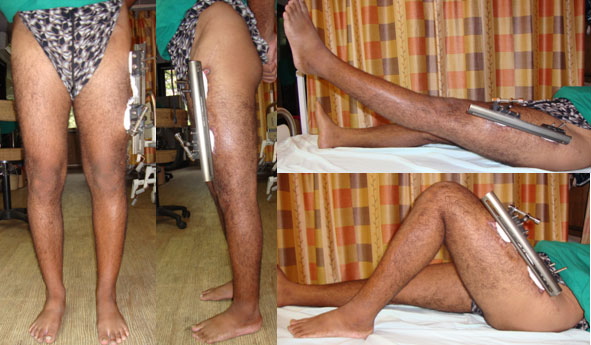
This 30 year old gentleman had met with an accident about 2 years back, following which there was a fracture of the left femur (thigh bone). He was treated at the local hospital where plating was done. The fracture healed and the plate was subsequently removed. He presented to us with deformity, shortening and awkward gait. The pictures here show the curvature in the thigh. The picture on the left shows an abnormal curvature of the thigh (red line) when we look from the front. The picture on the right shows the curvature of the thigh when we look from the side. Though it is a little proximal than normal, it is normal. |
|
| |
| |
| |
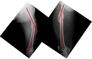
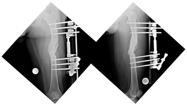
| The x-rays at presentation showing the angulations of the femur at the fracture site. The picture on the left is as seen from the front with red lines showing the axes of the upper and lower segments. The angle between the two axes was about 34°, which should be 0° normally. The picture on the right is from the side and shows the angle between the axes (red Lines) to be about 20°, which is about 12-14° normally. |
 |
|
| |
| |
| |
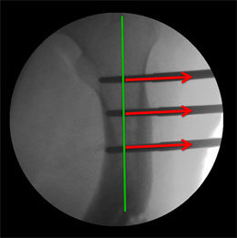 |
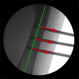
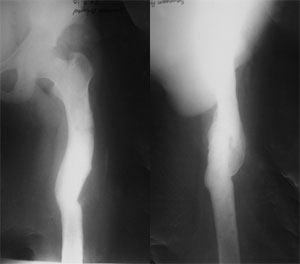
The C-arm picture during surgery, showing the axis of the proximal (upper) segment (green Line) and the red arrows depicting the angle of the Schanz screws applied. The screws are applied perpendicular to each segment, so that when the bone is cut and the screws are made parallel, the axes are aligned in a straight line automatically correcting the deformity. |
|
| |
| |
| |
| The C-arm picture during surgery, showing the axis of the distal (lower) segment (green Line) and the red arrows depicting the angle of the Schanz screws applied, should be perpendicular to the axis. |
 |
|
| |
| |
| |
 |
The pictures taken during surgery, showing the position of the Schanz screws and their orientation in different planes. |
|
| |
| |
| |
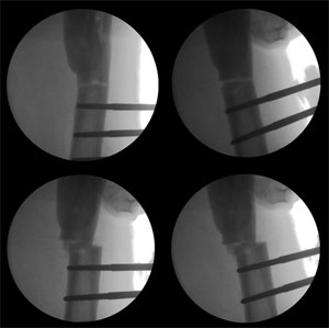
| The C-arm pictures of correction of the angulation and translation. Clockwise from top left –osteotomy above the lower group of screws, correction of angulation making the screws parallel, translation of the fragments relative to each other, final adjustment of angles and translation. |
 |
|
| |
| |
| |
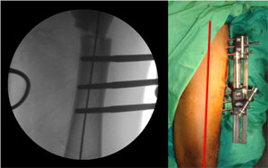 |
The C-arm picture on the left and the clinical picture showing the perfectly aligned axis. |
|
| |
| |
| |
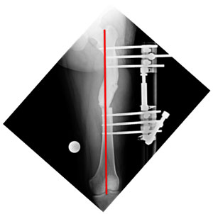
| The x-ray after surgery showing correction of axis. |
 |
|
| |
| |
| |
The clinical appearance and function just before the fixator was removed 6 months after surgery. Though the osteotomy had healed in 3 months but since the patient lives in another town he reported for fixator removal after 6 months.
|
| |
 |
| |
| |
| |
| The xrays just before fixator removal, showing complete healing of the osteotomy. |
| |
 |
| |
| |
| |
 |
The x-rays 1 month after fixator removal, showing healed screw holes. |
|
| |