- About Us
- Ilizarov Technique
- Process
- Case Studies
Fractures
• Acute Fracture - Comminuted Fracture Distal Humerus - Compound Fracture & VAC • Nonunion - Proximal Tibia - Tibia - Distal Humerus • Malunion - Femur Bone Deformity
• Acute Correction
- LRS - Bilateral Tibia - LRS - Rotational Deformity Femur(with Shortening) - LRS - Oblique Plane Deformity Femur - Nailing • Ilizarov Correction - Proximal Tibia - Distal Femur • Hexapod Correction - Metaphyseal Dysplasia Bone Infection
• Infection - Femur • Infected Nonunion - Proximal Femur Joint Problems (Arthritis)
• Hip Replacement • Knee Replacement • Intra Articular Osteotomy • High Tibial Osteotomy (HTO) • Arthrolysis - Elbow • Ilizarov Hip Reconstruction - FAQs
- Testimonials


Nonunion - Tibia
| At Presentation | ||
| 15 year old boy, presented with mobile nonunion of upper 1/3rd of tibia of 7 months duration. He had developed a pathological fracture secondary to osteomyelitis which was secondary to a previous surgery for hematoma evacuation. There was no sign of an active infection on presentation. | ||
 |
||
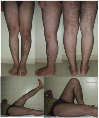
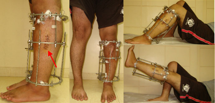
| Clinical Appearance at Presentation | ||
| The clinical pictures at presentation. The scars are from previous surgery. The bowing of left tibia was at the fracture site. Otherwise had full range of motion prior to surgery. | ||
 |
||
|
||
| 2 weeks post surgery | ||
| Clinical pictures at the end of 2 weeks showing good range of motion. The scar seen on the left picture is from the fibula was cut. | ||
 |
||
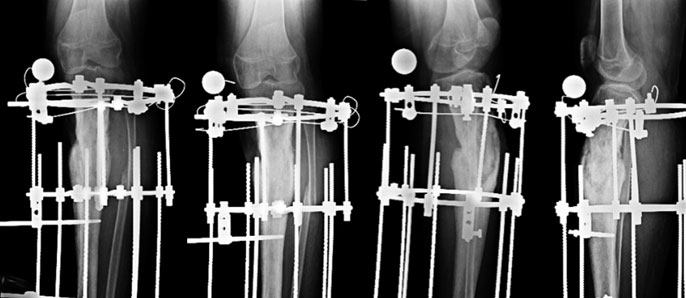
| 2 months | ||
| 2 months on, he continues to improve. The x-rays show signs of healing. There is formation of bridging callus. | ||
 |
||
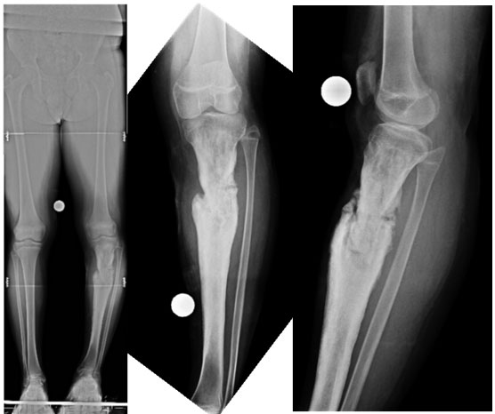
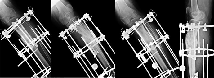
| 4 months post surgery – before fixator removal | ||
| At 4 months the fracture is healed. The callus is well consolidated. The fixator was then removed after gradual disassembly. | ||
 |
||
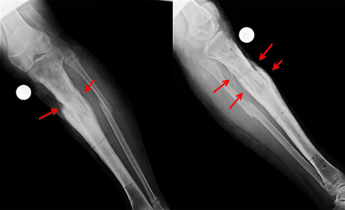
| After fixator removal | ||
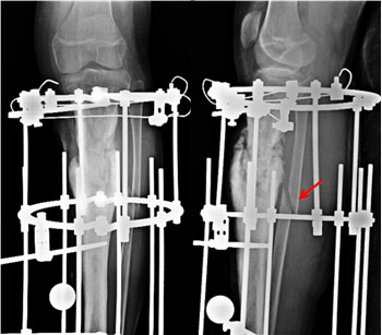
| The x-rays right after fixator removal. The callus is well seen (red arrow) bypassing &bridging the nonunion site. | ||
 |
||