- About Us
- Ilizarov Technique
- Process
- Case Studies
Fractures
• Acute Fracture - Comminuted Fracture Distal Humerus - Compound Fracture & VAC • Nonunion - Proximal Tibia - Tibia - Distal Humerus • Malunion - Femur Bone Deformity
• Acute Correction
- LRS - Bilateral Tibia - LRS - Rotational Deformity Femur(with Shortening) - LRS - Oblique Plane Deformity Femur - Nailing • Ilizarov Correction - Proximal Tibia - Distal Femur • Hexapod Correction - Metaphyseal Dysplasia Bone Infection
• Infection - Femur • Infected Nonunion - Proximal Femur Joint Problems (Arthritis)
• Hip Replacement • Knee Replacement • Intra Articular Osteotomy • High Tibial Osteotomy (HTO) • Arthrolysis - Elbow • Ilizarov Hip Reconstruction - FAQs
- Testimonials


Infected Nonunion Proximal Femur
|
||
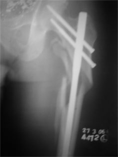
| 2006 | ||
| Unfortunately he developed infection and was re-operated during which an external fixator was applied to clear out the infection. | ||
|
||
|
||
|
||
|
||
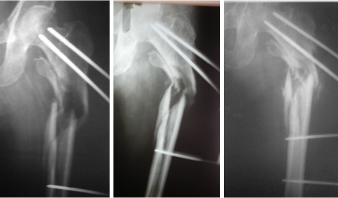
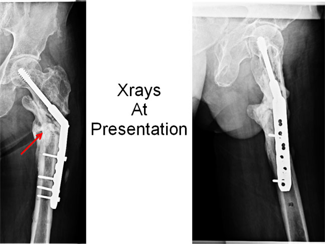
| Xrays At Presentation | ||
| The x-rays at presentation show loosening of screws, sclerosis at the fracture site, sequestrum (dead necrotic bone) (red arrow). | ||
 |
||
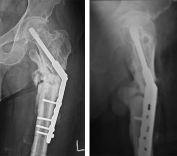
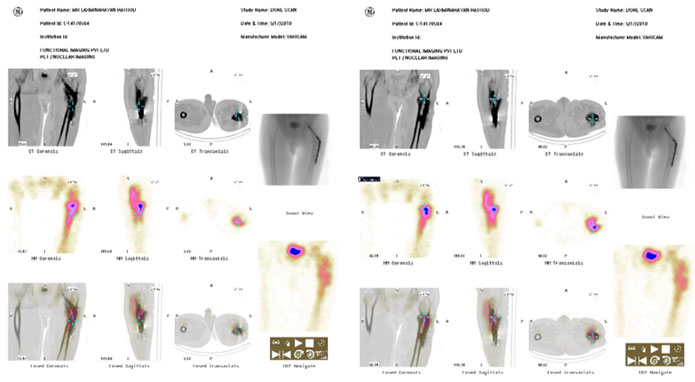
| The FDG-PET Scan showed infection right under the plate and in the antero-lateral aspect of the proximal femur. | ||
 |
||
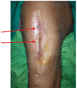
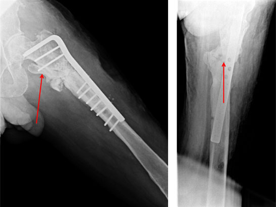
| The arrows show calcium sulfate pellets loaded with antibiotic. These pellets help in the elution of high dose of antibiotic locally to the infected area. This helps in better control of infection and obviates the need to give intravenous antibiotics for a long time (recommended period in osteomyelitis is 4-6 weeks of IV antibiotics). The advantage of using the calcium sulfate pellets is that it gets completely absorbed in around 6 weeks time, thereby preventing the need for another surgery. | ||
 |
||